
The “dog ear” is an infrequent but troublesome complication of wound closure. Asymmetric edges of a wound–which can be either iatrogenic, from a failure to align landmarks appropriately–or inevitable, from a traumatized wound with tissue loss and circular or asymmetric margins–leads to a closure conundrum in which one side of the wound is longer than the other. Closure in the usual fashion can result in puckering of the excess tissue and an unsightly cosmetic outcome.
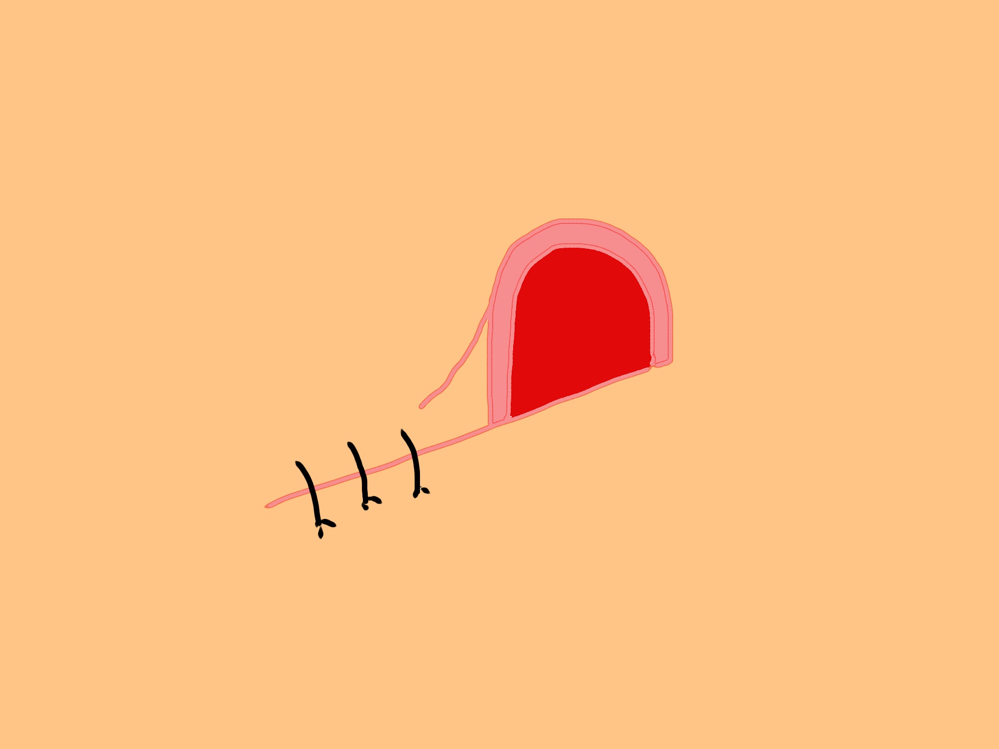 The “dog ear” is a deforming protrusion of a wound edge which can occur in the setting of irregular wound margins. This cartoon depicts a “half lying cone dog ear,” more commonly encountered in traumatic injuries than the “full cone” dog ear.
Previously on this site, I published a short video demonstrating a technique for correction of the dog ear. I advocated for this technique over others described in the literature based on its simplicity to perform, and thus easy adaptability for ED use. However, what I never liked about the technique was the requirement of wedge excision of tissue and lengthening of the scar, a step which is not familiar to most emergency physicians and urgent care providers.
As an alternative to this, I’d like to present another procedure, described this year in the Archives of Plastic Surgery. This novel technique is unique in that it closes the wound without sacrificing any tissue, a requirement of most dog ear correction techniques. The video above details each throw of the suture pattern.
The article also makes some great points about dog ear correction in general, which I’ll summarize in three sound “bites” below:
- “De-fatting” the subcutaneous tissue below a protruding dog ear can sometimes effectively allow the overlying skin to settle and lie symmetrically, precluding the need for any special suturing techniques.
- Sometimes, the best approach to a dog ear is to do nothing–treat it like you would treat any wound. Since the tissue protrusion is often due to local edema and worsened by infiltration of local anesthetics, some experts recommend that a dog ear with a height of less than 8 millimeters should be left alone.
- The technique above is recommended for dog ears with a projection from the skin surface no greater than 1/3 the length of its base (eg a 1:3 ratio), and not greater than an absolute projection of 15 mm from the skin surface.
