
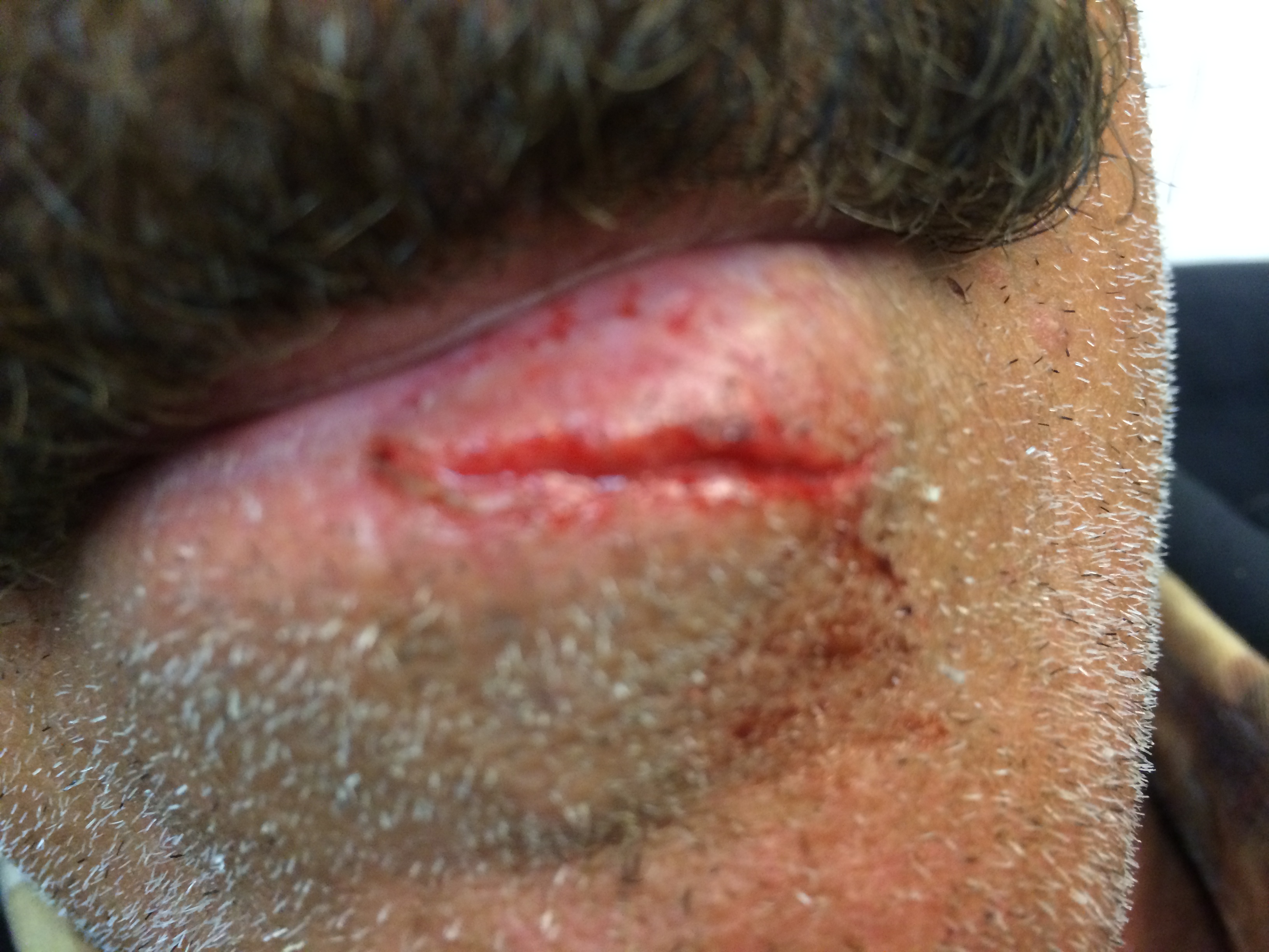
We spend all day looking at each other’s lips: listening to the words they form, interpreting the subtle social cues they convey, and sometimes imagining kissing them (when it’s the right person)! Thus, it’s natural that a patient and his/her friends and family will be preoccupied with the cosmetic closure of these wounds. It’s said that even a 1 millimeter “step-off” of the vermillion-cutaneous junction is quite noticeable and thus cosmetically unacceptable. So, let’s discuss how to make your next repair as elegant as possible.
In Part I of this post, we’ll discuss the evaluation and preparation of these wounds, which in truth is just as important to the repair as the suturing technique itself. In Part II, we’ll discuss the nuts and bolts of the suturing technique, including video of an actual repair.
Preparation: Anesthesia
Two ways that we distend and deform the margins of a wound are with anesthetic and irrigation. Undoubtedly, both are important practices. However, when cosmesis really matters, it pays to think about how we do it. I advocate for the use of a regional nerve block of the face rather than direct injection of local anesthetic in these situations.
Fortunately, every aspect of the upper and lower lips are amenable to nerve blocks!


Infraorbital nerve blocks provide excellent anesthesia to the ipsilateral half of the upper lip. (These blocks also cover the mid face including the skin of the nose, all the way up to the lower eyelid.) A mental nerve block is a great technique to achieve anesthesia of the ipsilateral half of the lower lip. (These blocks also cover the skin of the face down to the angle of the mandible.)
Preparation: Irrigation
Decontamination of traumatic lacerations through irrigation is a general maxim of wound care. There is much discussion in the literature regarding the optimal type of irritant fluid, delivery system to optimize pressure, and volume used. Some of the major points and conclusions of said discussions are highlighted elsewhere on this site. In general, high volume and high pressure of irrigation are what we are used to teaching…
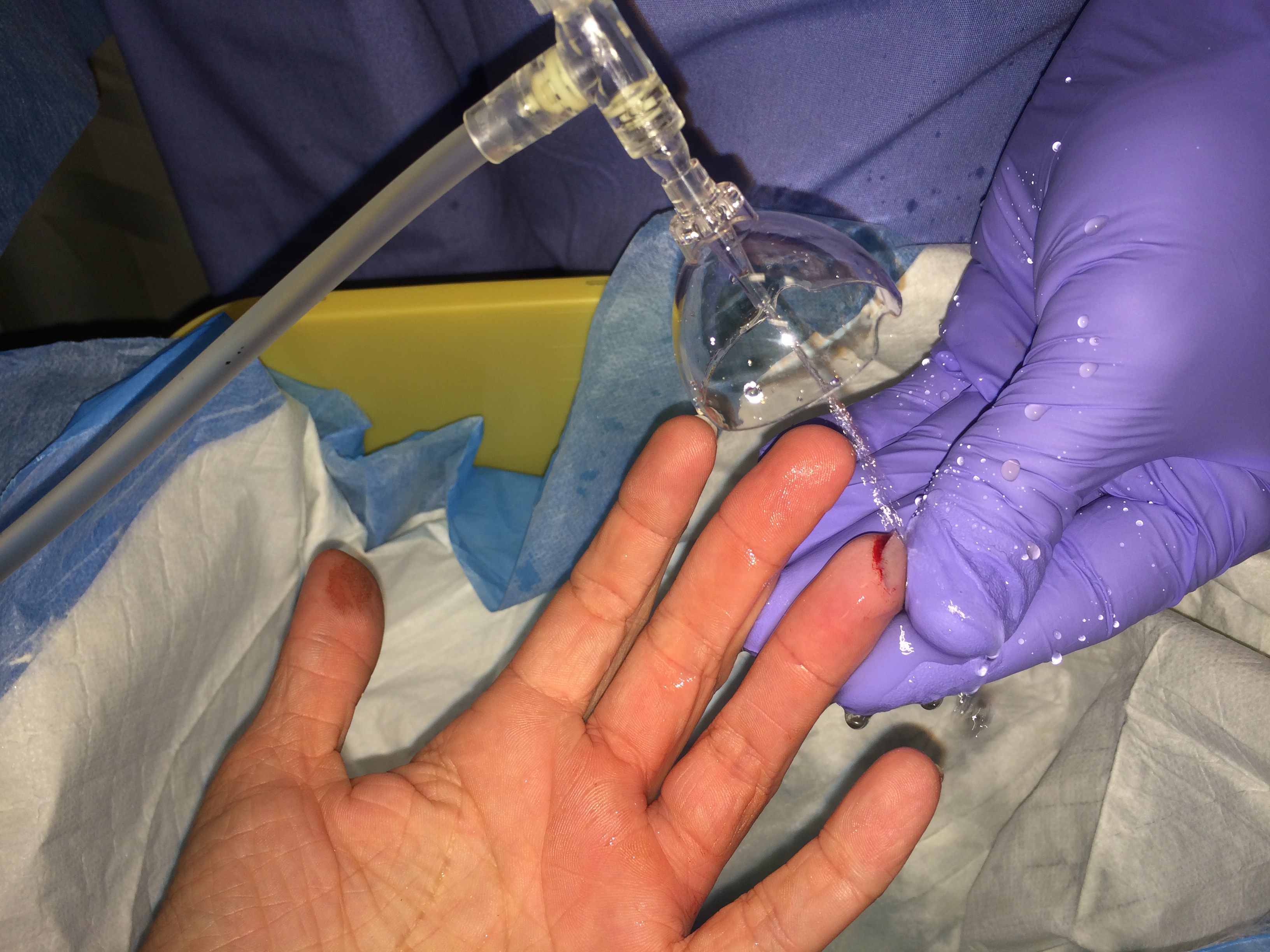
However, this may not hold true for wounds of the face.
One important downside of high volume irrigation at high pressure is dissection through tissue planes. This leads to distension of the subcutaneous tissues and distortion of wound margins. If this is a necessary evil in order to prevent infection, so be it, but there is at least some data to suggest that maybe it’s not. A 1998 study published in the Annals of Emergency Medicine suggests that for clean wounds of the face and scalp, irrigation (or lack thereof) does not affect rate of infection nor cosmetic outcome. Definitely some food for thought–experienced practitioners, when is the last time you saw an infection of an uncomplicated scalp or facial laceration? I think this paper makes a reasonable argument against copious irrigation of selected clean facial wounds, and I’ve started to consciously skip irrigation on facial wounds when I think the wound margin distension may effect closure. The lip is the perfect example of this.
Preparation: Exploration
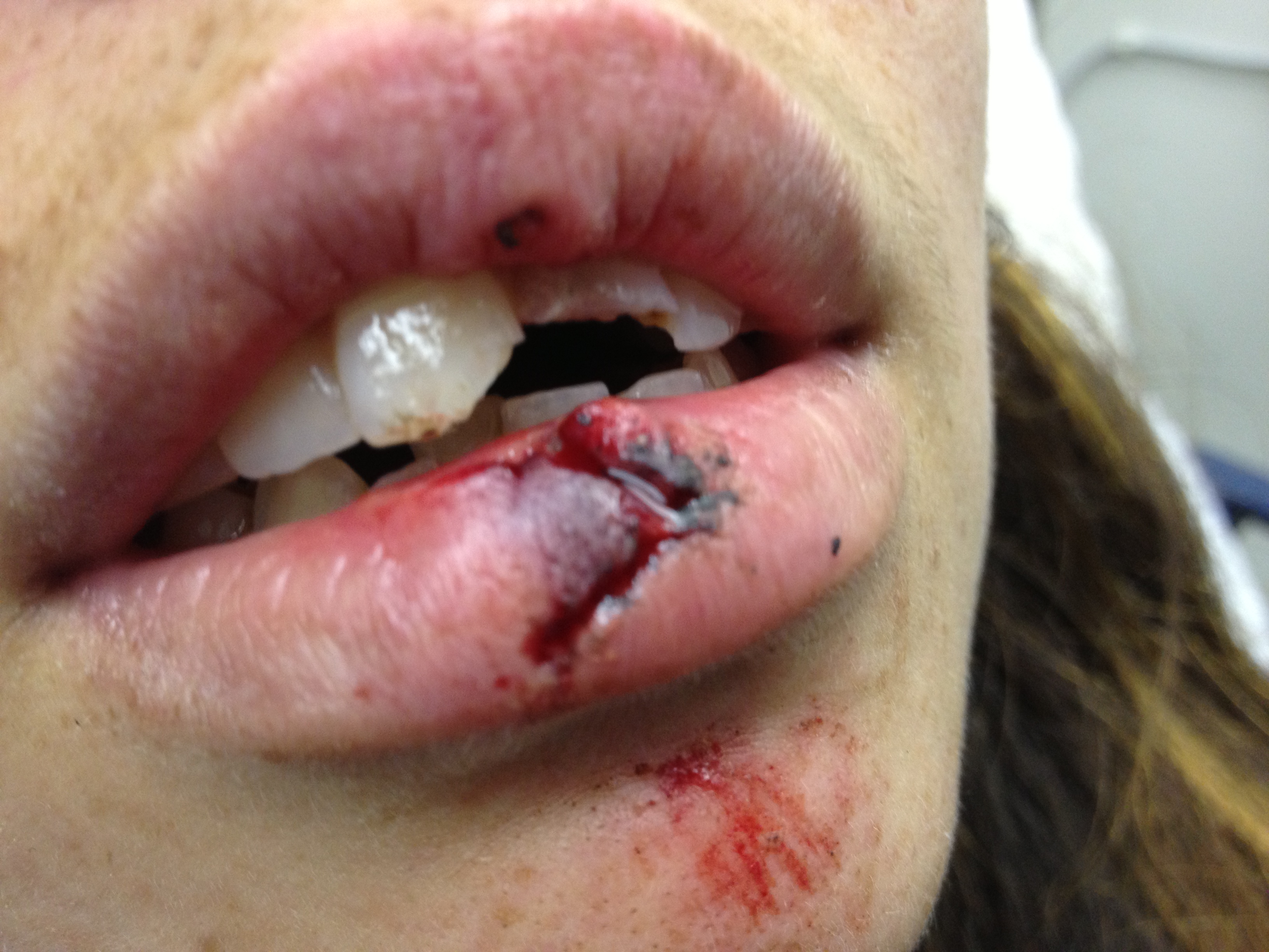 When teeth are chipped (often the case with lip lacerations), ensure no fragments remain embedded in the wound.
Lip lacerations often go hand-in-hand with injuries to the teeth. (Actually, the teeth are often the cause of the lacerations!) It’s very important to remember to explore that wound carefully before closure, especially if a chipped tooth is present and the missing fragments are not otherwise located. If these tooth fragments remain embedded in a wound, they may act as foreign bodies which can be a nidus for infection. Soft tissue radiographs can visualize small tooth fragments if there is any doubt.
Great! Your wound is elegantly anesthetized, judiciously cleansed, and meticulously explored. Now, let’s get on with the repair.

One thought on “Lip Lacerations, Part I”
Comments are closed.